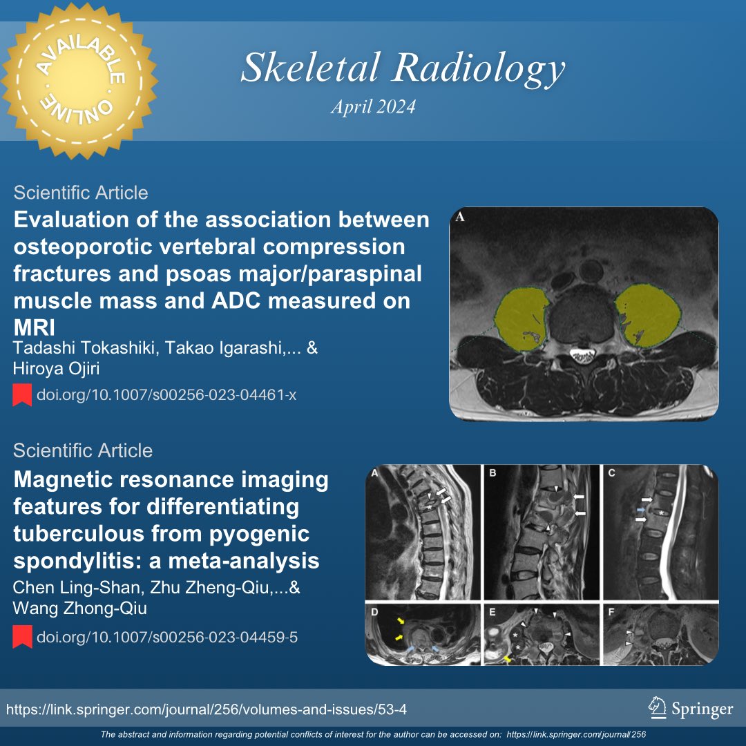
Access the April issue of the Skeletal Radiology Journal
To read the full articles, use the following links:
🔴 rdcu.be/dE8NO
🔴 rdcu.be/dE8PW
#SkeletalRadiology #SkeletalJournal #MSKrad #orthopedics #radres #pathology
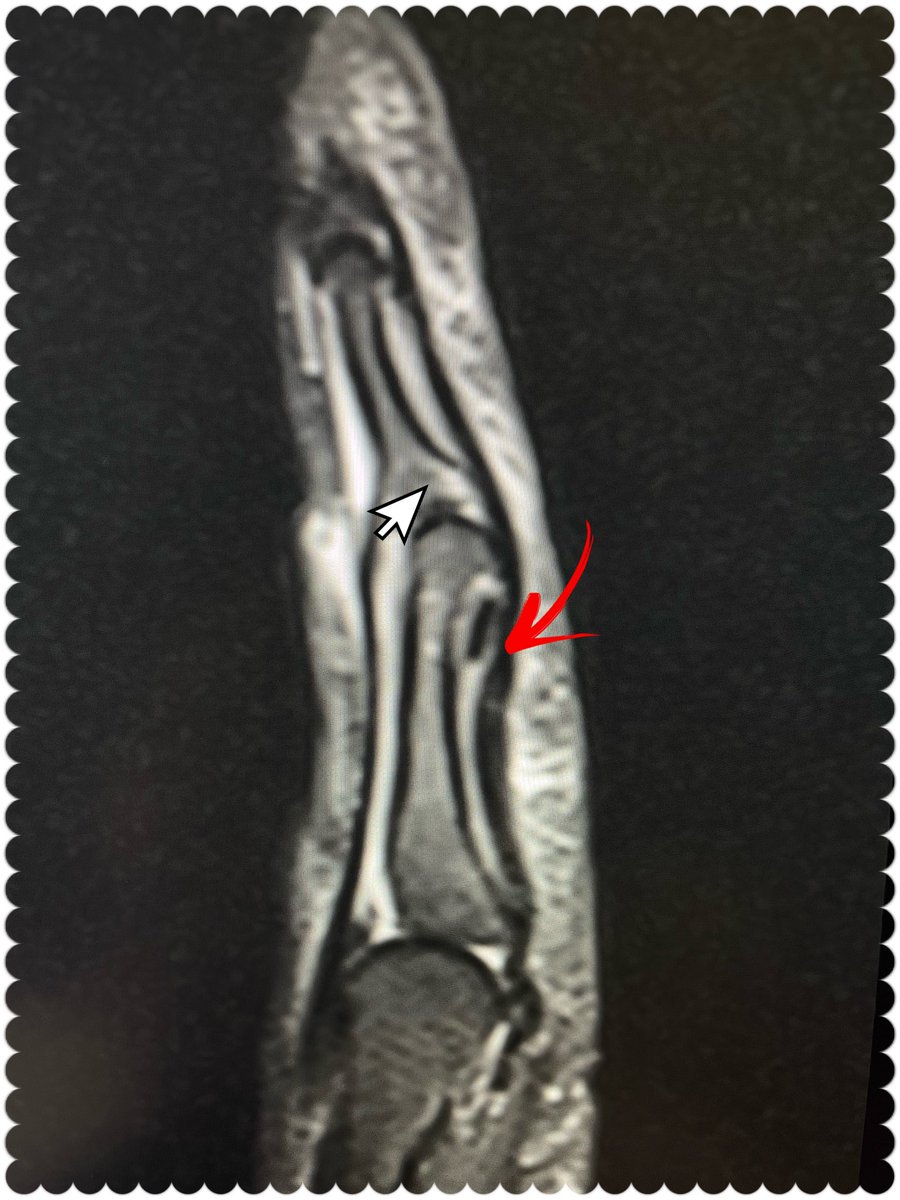
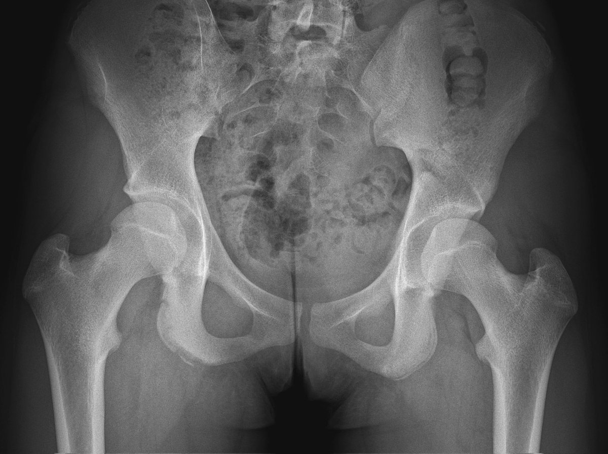
#CaseOfTheWeek ‼️🥳‼️
☢️🩻☠️Case#16☠️🩻☢️
👁️test👁️
📲➡️➡️ #Diagnosis ❔❓❔
#FOAMRad #RadEd #MedEd #OrthoEd #OrthoTwitter SSR_RWG UWisconsin Radiology Residents POSNA

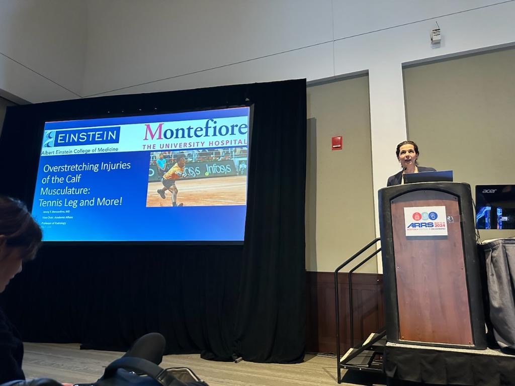
As always a pleasure to participate as speaker faculty at #ARRS2024 talking on #MRI #muscle alongside amazing #MSKRad colleagues:Miriam Bredella, MD, MBA, FACR Meghan Jardon Atul K. Taneja, MD, PhD Flavio Duarte Silva
Thank you Avneesh Chhabra, MD, MBA, FACR for organizing! ARRS

Back at Erasmus MC combining clinical work and research into PET/MRI for musculoskeletal diseases after two years as visiting Ass Prof UW Radiology UW MSK Imaging & Intervention. Had an amazing time with special thanks to Scott Reeder, Kenneth S. Lee, Pain Imaging Lab, David Harris and Ali Pirasteh!

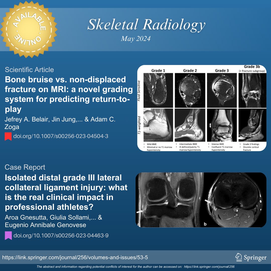
Access the May issue of the Skeletal Radiology Journal
To read the full articles, use the following links:
🔴 rdcu.be/dD7Om
🟣 rdcu.be/dE84A
#SkeletalRadiology #SkeletalJournal #SKRAJournal #MSKrad #sportsmedicine

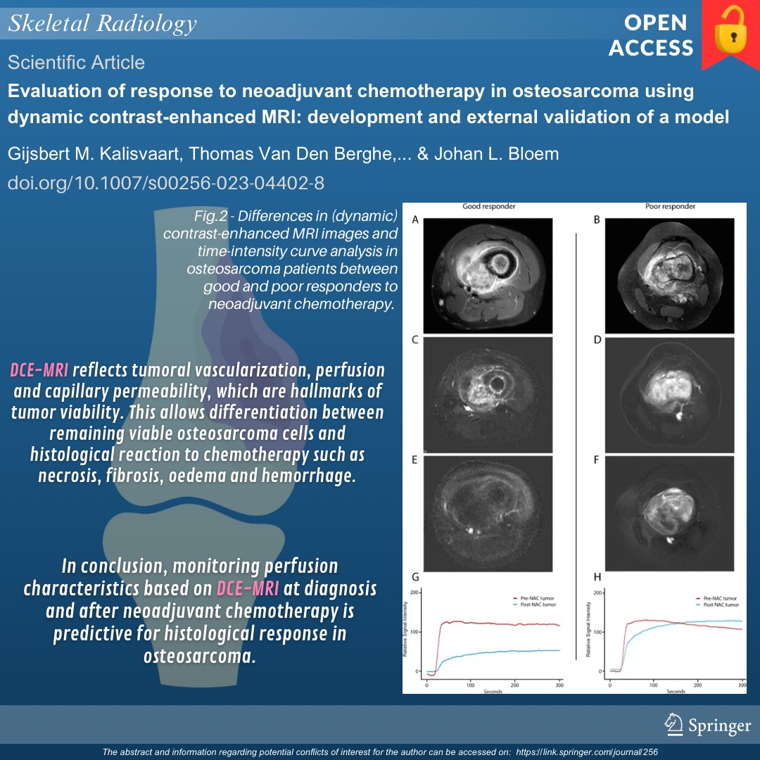
Check out the new infographic:
🔴 Evaluation of response to neoadjuvant chemotherapy in osteosarcoma using dynamic contrast-enhanced MRI: development and external validation of a model
🔓Open access 👉 doi.org/10.1007/s00256…
#MSKRad #SkeletalRad #orthotwitter #oncology

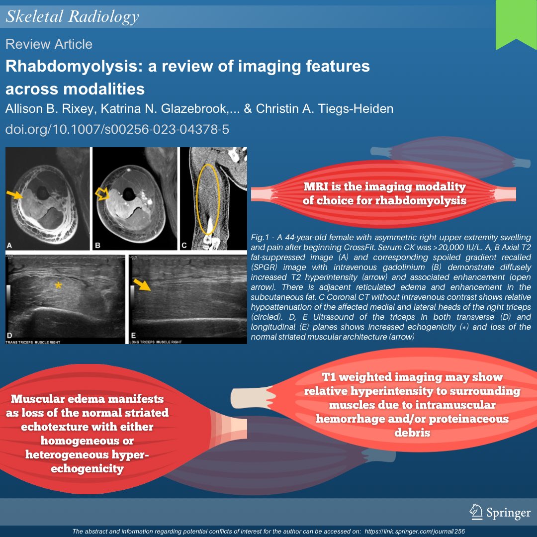
Check out our informative infographic:
🟢 Rhabdomyolysis: a review of imaging features across modalities | rdcu.be/dwgJZ
#SkeletalRadiology #SkeletalJournal #SKRAJournal #MSKrad #orthopedics


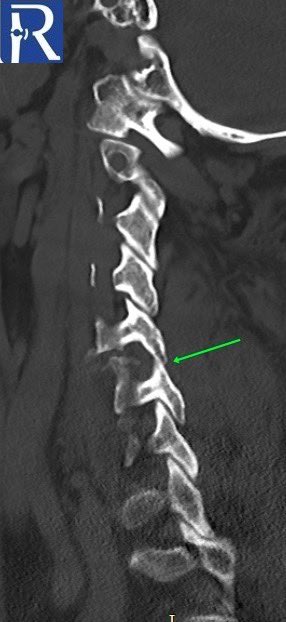
A 73-year-old female passenger was admitted following a loss of consciousness after a high-speed motor vehicle accident. #radiology #mskrad iology #mskrad #radtwitter #radres #medstudent #neurosurgery #neuroradiology #MedTwitter #emergencymedicine #Neurology #radres #neurores


Bone Age Prediction under Stress doi.org/10.1148/ryai.2… Shahriar Faghani MD MayoAILab Brad Erickson #MSKRad #BoneAge #MachineLearning


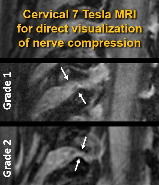
Out now in print & #OpenAccess : 7T MRI of the cervical neuroforamen 👉doi.org/10.1097/rli.00…
We demonstrated that #7T DESS imaging can directly assess cervical nerve root compression, pushing the boundaries of spine imaging
Georg Feuerriegel Balgrist University Hospital Siemens Healthineers Press #MSKrad


. Additionally, there is mild associated rotatory subluxation without dislocation of the left facet joint (indicated by the green arrow). #radiology #mskrad #radtwitter #neurosurgery #Emergencyradiology #radres
