Lea Alhilali, MD(@teachplaygrub) 's Twitter Profileg
Lea Alhilali, MD

@teachplaygrub

Neuroradiologist @HRInstitute_AZ. @BarrowNeuro. Striving to make learning neuroimaging and anatomy fun. If I can make you laugh, I can make you learn.

ID:1519179930923266048

calendar_today27-04-2022 05:01:44

5,2K Tweets

52,6K Followers

165 Following

Lea Alhilali, MD(@teachplaygrub) 's Twitter Profile Photo

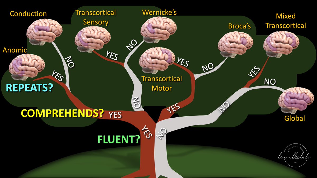
When you’re asked to localize the lesion in a patient with aphasia, do you suddenly feel speechless?

At a loss for words to categorize the type of aphasia?

Never fear—here is the decision tree for patients w/aphasia and the associated anatomic correlates

Three main questions:

When you’re asked to localize the lesion in a patient with aphasia, do you suddenly feel speechless? At a loss for words to categorize the type of aphasia? Never fear—here is the decision tree for patients w/aphasia and the associated anatomic correlates Three main questions:
account_circle
Lea Alhilali, MD(@teachplaygrub) 's Twitter Profile Photo

If you are only looking at the optic nerve on Orbital MRIs, you need to open your EYES!

Don’t be blind to all the other nerves in the orbit!

Branches of trigeminal V1 &V2 and the oculomotor nerve all can cause pathology in the orbit

But if you don’t look, you won’t see!

Let

account_circle
Lea Alhilali, MD(@teachplaygrub) 's Twitter Profile Photo

Are you right when it’s bright?

Bright cortical signal on diffusion images is classically associated w/hypoxic-ischemic injury.

But there are many mimics!

Do you know how to recognize the different patterns of cortical restricted diffusion? Here’s a figure & some pearls to

account_circle
Lea Alhilali, MD(@teachplaygrub) 's Twitter Profile Photo

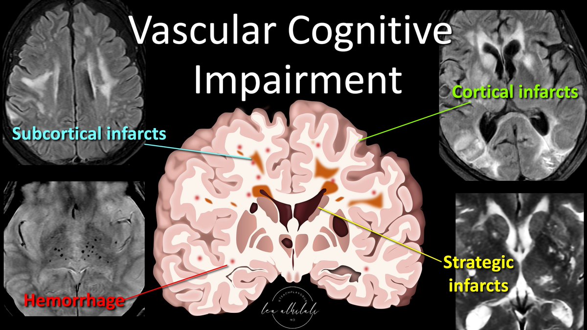
1/Having trouble remembering what you should look for in vascular dementia on imaging?

Almost everyone worked up for dementia has infarcts. Which ones are important?

Here’s a thread on the key findings in vascular dementia!

1/Having trouble remembering what you should look for in vascular dementia on imaging? Almost everyone worked up for dementia has infarcts. Which ones are important? Here’s a thread on the key findings in vascular dementia!
account_circle